Basics for performing a high-quality color Doppler sonography of the vascular access
Mario Meola , Jose Ibeas , Gianfranco Lasalle and Ilaria Petrucci
Abstract
In the last years, the systematic use of ultrasound mapping of the upper limb vascular network before the arteriovenous fistula (AVF) implantation, access maturation, and clinical management of late complications is widespread and expanding.
Therefore, a good knowledge of theoretical outlines, instrumentation, and operative settings is undoubtedly required for a thorough examination. In this review, the essential Doppler parameters, B-Mode setting, and Doppler applications are considered. Basic concepts on the Doppler shift equation, angle correction, settings on pulse repetition frequency, operative Doppler frequency, gain are reported to ensure adequate and correct sampling of blood flow velocity. A
brief analysis of the Doppler inherent artefacts (as random noise, blooming, aliasing, and motion artefacts) and the adjustment setting to minimize or eliminate the confounding artefacts are also considered. Doppler aliasing occurs when
the pulse repetition frequency is set too low. This artefact is particularly frequent in vascular access sampling due to the high velocities range registered in the fistula’s different segments. Aliasing should be recognized because its correction
is crucial to analyse the Doppler signals correctly. Recent advances in instrumentation are also considered about a potential purchase of a portable ultrasound machine or a top-of-line, high-end, or mid-range ultrasound system.
Last, the pulse wave Doppler setting for vascular access B-Mode and Doppler assessment is summarized.
Keywords
B-mode ultrasound, pulse wave Doppler, arterio-venous fistula, Doppler parameters, Doppler/B-mode setting
Date received: 10 April 2021; accepted: 22 April 2021
Introduction
In the last years, the routine use of ultrasound in the map– ping of upper limb vascular network before the arterio– venous fistula (AVF) implantation, access maturation, and clinical management of late complications is widespread
and growing. Therefore, a good knowledge of theoretical outlines, instrumentation, and operative settings is undoubtedly required for a thorough examination.
Ultrasound, similar to MRI, is a “multiparametric” imaging technique because it analyses and represents native echo-signals originating from tissues and vessels in
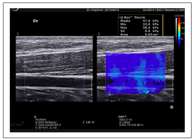
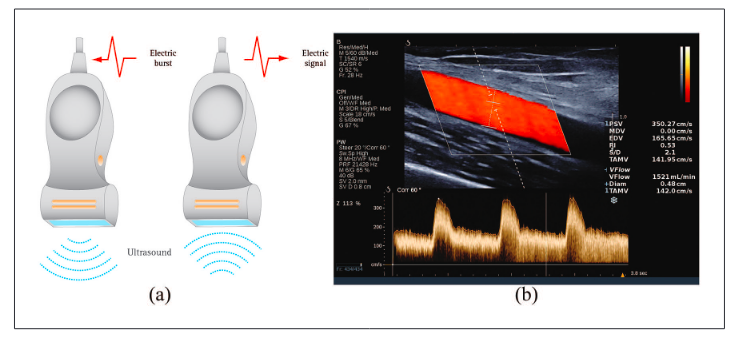
different ways.1 B-Mode represents structural echoes as brightness dots and generates a grey scale image of the tissue. Doppler-mode imaging analyses in a region of interest (ROI) the backscattering from the red blood cells (RBCs) moving in the vessels. (a) Color Doppler represents the blood flow-velocity changes as a dynamic mapping where the flows move towards or away from the transducer and are respectively depicted with red and blue…
1Sant’Anna School of Advanced Studies, Pisa, Italy
2Consorcio Corporacion Sanitaria Parc Tauli, Sabadell, Barcelona, Spain
Corresponding author:
Mario Meola, Sant’Anna School of Advanced Studies, Piazza Martiri
della Libertà, 33, Pisa 56127, Italy.
Emails: m.meola@santannapisa.it; mario.meola@unipi.i