Advances in vascular anatomy and pathophysiology using high resolution and multiparametric sonography
Petrucci Ilaria , Meola Mario and Fiorina Ilaria
Abstract
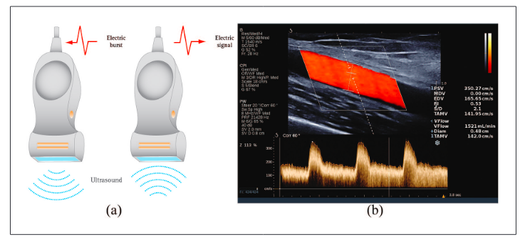
B-mode and Color Doppler are the first-line imaging modalities in cardiovascular diseases. However, conventional ultrasound (US) provides a lower spatial and temporal resolution (70–100 frames per second) compared to ultrafast technology which acquires several thousand frames per second. Consequently, the multiparametric ultrafast platforms manage new imaging algorithms as high frequency ultrasound, contrast-enhanced ultrasound, shear wave elastography, vector flow, and local pulse wave imaging. These advances allow better ultrasound performances, more detailed blood flow visualization and vessel walls’ characterization, and many future applications for vascular viscoelastic properties
evaluation.
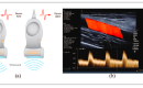
In this paper, we provide an overview of each new technique’s principles and concepts and the real or potential applications of these modalities on the study of the artery and venous anatomy and pathophysiology of the upper limb before and after creating a native or prosthetic arterio-venous fistula. In particular, we focus on high-frequency ultrasound that could predict cannulation readiness and its potential role in the venous valvular status evaluation before vascular access creation; on contrast-enhanced ultrasound that could improve the peri-operative imaging evaluation during US-guided angioplasty; on shear wave elastography and local pulse wave imaging that could evaluate preoperative vessels stiffness and their potential predictive role in vascular access failure; on vector flow imaging that could better characterize the different components of the vascular access complex flow.
Keywords
Multiparametric ultrasound, shear wave elastography, high-frequency ultrasound, vector flow imaging, local pulse wave velocity, vascular access
Date received: 19 June 2020; accepted: 6 May 2021
Introduction
Ultrasound (US) is the first-line imaging modality for screening, diagnosis, and monitoring treatment in cardio- circulatory pathology because of its safeness, non-invasiveness, wide availability, and low cost. Conventional ultrasound modalities as B-mode, color Doppler, and spectral analysis allow the recognition of vessel wall and bloodstream changes, depicting the site of vascular stenoses and occlusions.1,2 Consequently, US plays a pivotal role also in hemodialysis vascular access creation and surveillance.
Conventional platforms have a limited frame rate (images/second) because they use the line-by-line scanning acquisition method. This technical approach provides a limited temporal resolution. Recent ultrafast platforms acquire mage information at frame rates of several thousand Hz, while conventional ultrasound systems acquire images at only 70–100 frames per second. The higher temporal and
spatial resolution of this technical approach allows managing all the new imaging algorithms as high-frequency.
1Institute of Life Sciences, S. Anna School of Advanced Studies, Pisa,
Italy
2Institute of Life Sciences, S. Anna School of Advanced Studies,
Department of Internal Medicine, University of Pisa, Pisa, Italy
3Radiodiagnostic and Interventional Radiology Department, Fondazione
IRCCS Policlinico San Matteo, Pavia, Italy
Corresponding author:
Petrucci Ilaria, Institute of Life Sciences, S. Anna School of Advanced
Studies, Piazza Martiri della Libertà, 33, Pisa 56127, Italy.
Email: i.petrucci@santannapisa.it